Crystallography

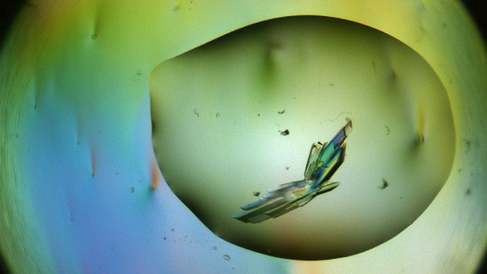
X-ray crystallography produces high-resolution models of proteins to allow an understanding of their structure and function at the atomic level. The process begins by growing a crystal of the protein of interest that contains millions of copies of that protein arranged in an ordered and regularly-repeating fashion. When an X-ray beam is directed through these crystals, electrons within the ordered proteins diffract the X-rays in an interpretable manner. Through analysing the pattern of diffraction the electron density within the protein of interest can be elucidated, which is then used to create a model of the protein's structure. These structures give scientists essential insights into human health and disease. For example, X-ray crystallography is vital in modern drug discovery as it allows the molecular interactions between drugs and their targets to be visualised for efficient drug optimisation and to reveal the drug's mode of action.
The Crystallography Facility provides access to the latest state-of-the-art robotics for performing high-throughput macromolecular crystal structure determination. These include mosquito crystal, dragonfly crystal, tecan and formulatrix rock imager systems, which combine together to allow efficient screening of thousands of crystallisation conditions to identify initial crystal hits and optimise them into usable crystals. The Facility has well established and regular access to various synchrotrons at the Diamond Light Source, and elsewhere, for subsequent rapid determination of high-resolution structures.
