The Dupree Group and colleagues from the University of Warwick have published a new paper in Nature Plants on the interaction between carbohydrate chains in the plant secondary cell wall.
Plant cell walls consist of multiple layers: the middle lamella, a pectin-rich layer forming the interface between plant cells to hold them together; the primary cell wall of cellulose, hemicellulose and pectin, which provides strength and flexibility for growth; and the secondary cell wall, a thick layer formed inside the primary cell wall in some cell types. This secondary cell wall confers cells with added strength and support through its abundance of xylan and cellulose carbohydrate chains.
To create the secondary cell wall, long chains of xylan sugar units have to bind to cellulose. It was recently shown that this occurs through xylan chains adopting a ribbon- or screw-like structure, where every second xylan unit faces in the same direction.1 Interestingly, every second xylan unit is also decorated with an acetate or glucuronic acid group, meaning these additions are all found on the same xylan face. It was therefore previously proposed that this even-specific arrangement of xylan decorations may allow xylan to bind the hydrophilic (water-facing) surface of cellulose.2-4
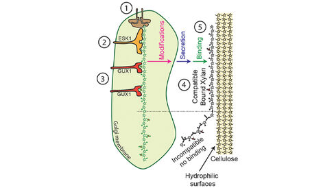
In their new paper, the Dupree Group use mass spectrometry to investigate xylan decorations from Arabidopsis with a mutant form of ESK1 (an enzyme responsible for adding acetate groups to xylan); finding that ESK1's normal function is important in generating the even pattern of xylan acetate decorations. Furthermore, the ESK1 mutant profoundly alters the even pattern of xylan glucuronic acid decorations normally placed via the enzyme GUX1. Subsequent NMR spectroscopy analyses of xylan chains from ESK1 mutant plants show that loss of even-specific xylan decorations cause the chains to adopt an abnormal screw-like structure that cannot properly interact with cellulose and which is more mobile within the cell wall. The findings from the Dupree Group suggest the following model for the binding of xylan to cellulose:
- Xylan chains are created in the Golgi apparatus.
- ESK1 adds acetate groups to every second xylan sugar unit within the xylan chain.
- GUX1 adds glucuronic acid groups to even-spaced xylan units, based on the pattern of acetate groups added by ESK1.
- The xylan chain adopts a screw-like structure where acetate and glucuronic acid groups on even-spaced xylan units are aligned on the same face of the chain.
- The xylan chain binds the hydrophilic surface of cellulose.
Importantly, biofuel processing, paper manufacture and digestion of feed all require the separation of xylan from cellulose. This new model for the binding of xylan to cellulose has increased our understanding of the structure of the secondary cell wall, which may lead to new methods for the preparation and application of biomaterials from plants in the future.
1Simmons et al., Nat. Commun. 7:13902, 2016.
2Bromley et al., Plant J. 74:423, 2013.
3Busse-Wicher et al., Plant J. 79:492, 2014.
4Busse-Wicher et al., Plant Physiol. 171:2418, 2016.
