Structures
Here are the structures that we and our lab have solved. All figures were made with Molscript and rendered with Raster3D. Click the image to take you to the Proteopedia page in a new tab.
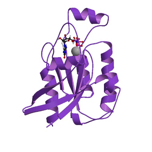 |
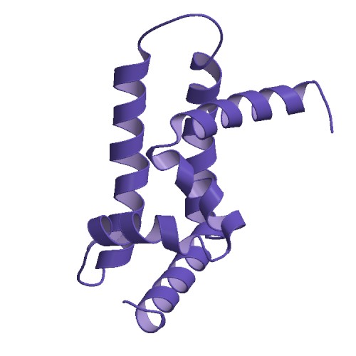
Cdc42 GMPPNP-bound (never deposited in the PDB). Solved in 1997-1998. The magnesium ion is shown as a spacefill and the nucleotide as a ball and stick representation. |
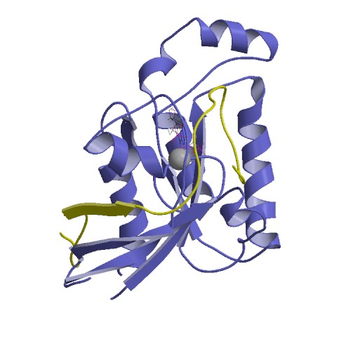 |
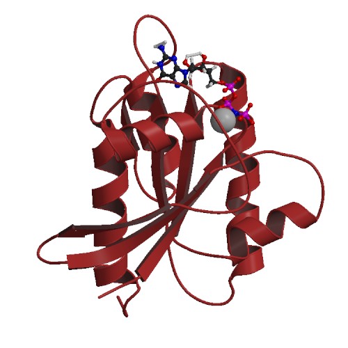
Cdc42 bound to GMPPNP and in complex with the G protein binding domain of the non-receptor tyrosine kinase ACK1. This structure was published in 1999. Cdc42 is shown in blue and the ACK is in yellow. The pdb code for this structure is 1cf4. |
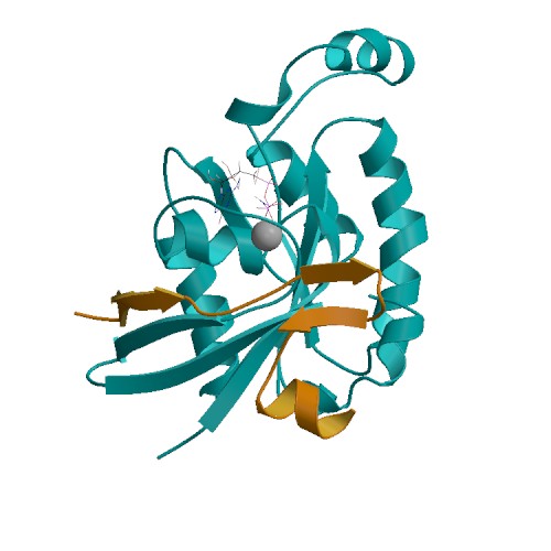 |
Cdc42 bound to GMPPNP and in complex with the G protein binding domain of the serine/threonine kinase PAK. This structure was published in 2000. Cdc42 is shown in cyan and PAK is in orange. The pdb code for this structure is 1e0a. |
 |
The Ral binding domain from the exocyst component protein Sec5. This structure was published in 2003. The pdb code for this structure is 1hk6. |
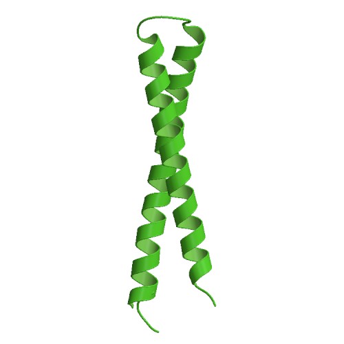 |
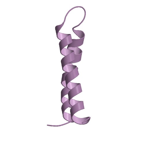
The HR1b domain from the serine/threonine kinase PRK1 (protein kinase C related kinase 1). This structure was published in 2003. The pdb code for this structure is 1urf. |
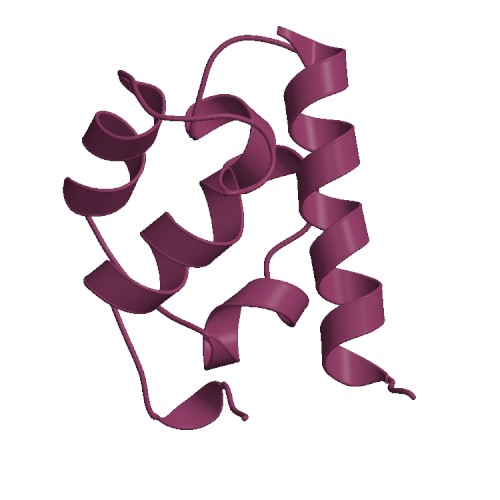 |
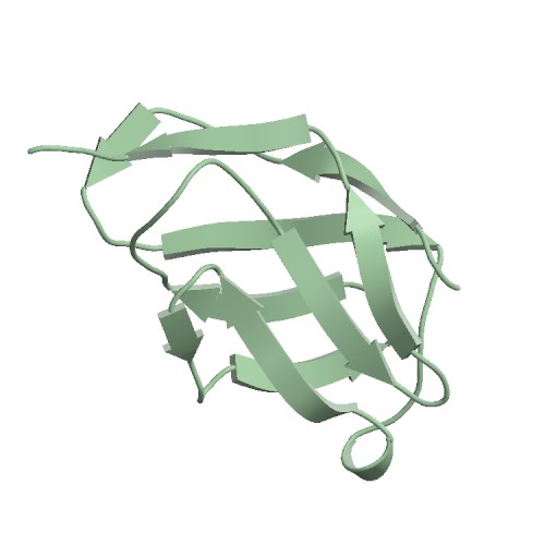
The structure of the SAM (sterile alpha motif) domain from the yeast signalling protein Ste50. This structure was published in 2004. The pdb code for this structure is 1uqv. |
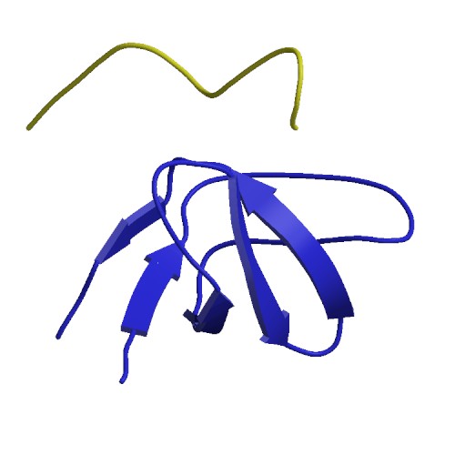 |
The structure of the SH3 domain from the Cdc42/Rac exchange factor b-PIX in complex with a peptide from the serine/threonine kinase a-PAK. This structure was published in 2005. The pdb code for this structure is 1zsg. |
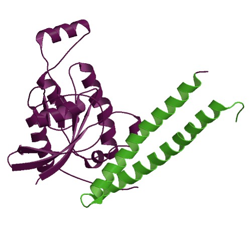 |
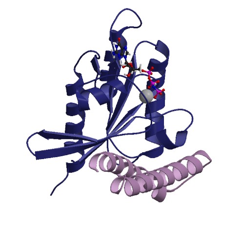
The structure of the HR1b domain from the serine/threonine kinase PRK1 in complex with the small G protein Rac1 bound to GMPPCP. This structure was published in 2008. The pdb code for this structure is 2rmk. |
 |
The structure of the Arl2 effector BART. This structure was published in 2009. The pdb code for this structure is 2k9a. |
 |
The structure of the small G protein RalB bound to GMPPNP. This structure was published in 2009. The pdb code for this structure is 2ke5. |
 |
The structure of the Ral binding domain of RLIP76 (RalBP1). This structure was published in 2010. The pdb code for this structure is 2kwh. |
 |
The structure of the Ral binding domain of RLIP76 (RalBP1) in complex with the small G protein RalB. This structure was published in 2010. The pdb code for this structure is 2kwi. |